
Inspired by PA4Ortho, I submit the following:
Allow me to briefly don the cape of Captain Obvious, and state that Xray machines are somewhat hard to come by out in the field.
However, there is a technique to detect long bone fractures in the field with a stethoscope and tuning fork, absent the gold standard X-ray.
It is published here, and the technique is as follows:
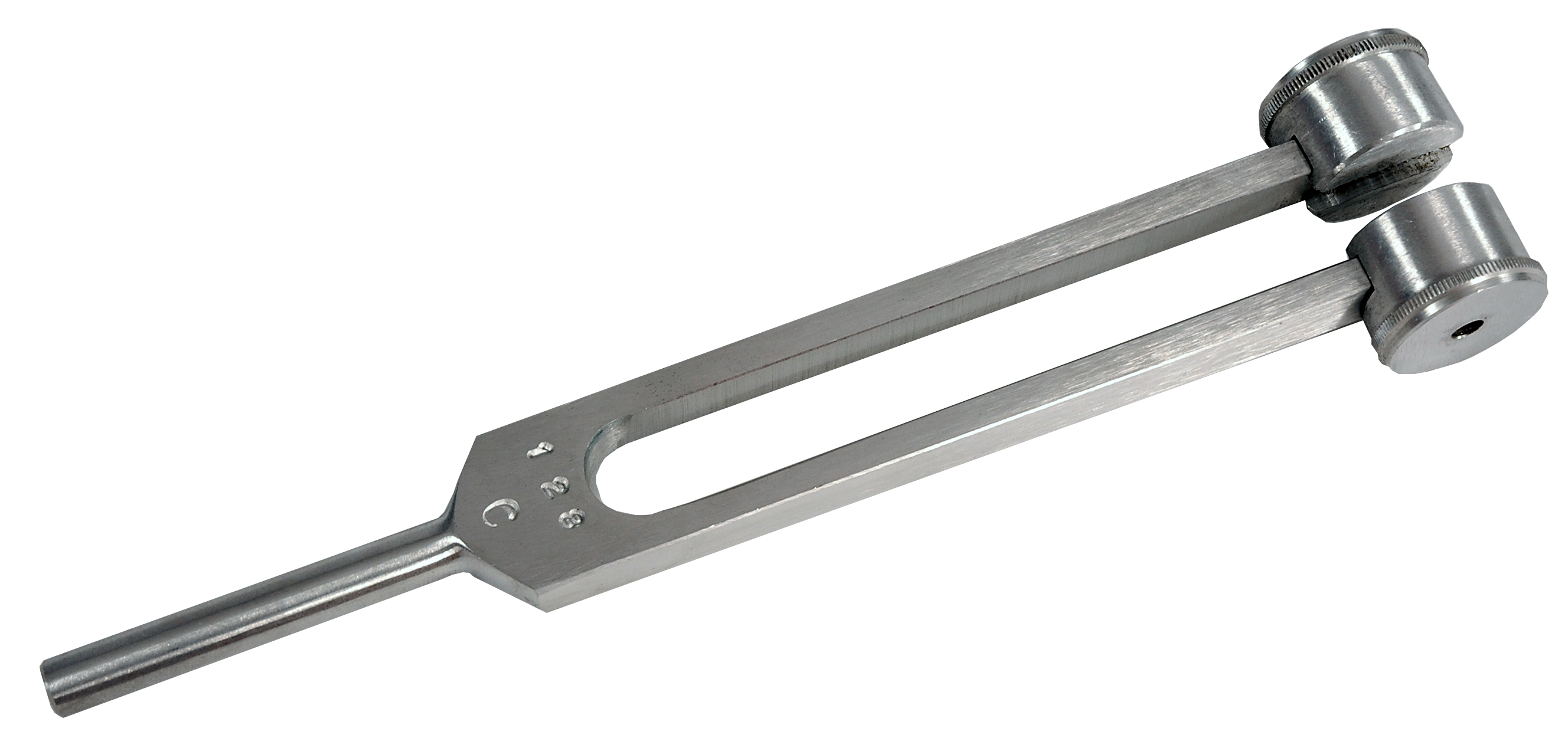
Place your stethoscope and your tuning fork ( a 128 Hz model was used, but I suspect you could use a 256 Hz model) on opposite sides of the location of the suspected break. Compare to the uninjured side. You should hear a clear transmission of the sound of the tuning fork on unbroken bones, and it should be dampened on the site of injury.
In the publication above, this was about 80% sensitive (picked up 10 out of 12 that had a fracture) and 80% specific (correctly ruled out 20 of 25 who did not have a fracture).
See here for related video:
My experience with this is that occasionally the vibration of the tuning fork caused increased pain at the site of the break; the article above claims this is “painless”. My patients never complained of dramatically increased pain, just noticeable increased pain, so I did not feel as if I tortured them. My hypothesis is that causes the broken ends of the bone to vibrate against one another, and this caused worsening pain at the fracture site. It has no effect on unbroken bones.
I’ve not formally tested or published that last bit.
I know, I know, I’m breaking my rule about citing data. But there it is.
Assignment #1: find a tuning fork (they are very cheap on Amazon, and much cheaper than an Xray machine) and actually do this. Practice so that you know what normal sounds like; afterwards, abnormal is quite obvious.
–Grouch
Update: Updated to better reflect the article quoted. Also, the references from the article cite several other papers that also investigated this technique, which you can review at your leisure.

Pingback: Hogwarts: The Poor Man’s X-Ray | Western Rifle Shooters Association·
Pingback: Field Medical Assessment | Head Space·
Doc Grouch hat tip, well done.
Most fractures can be picked up on by the usual pain swelling or deformity issues. Palpate gently along the bone feeling for pain or step off, percuss gently then firmly to find the point of most pain. If in doubt splint it for 3-10 days and make it non wt bearing*, if its pain free then it was likely not broken or is reasonably stable, gentle progression of activities as tolerated avoiding impact for a few more weeks. If it still hurts 3-10 days later then splint cast or protect it for 6 weeks on average. Different bones take different time frames to heal.
The tuning fork or ‘ultrasonic’ tooth brush are good adjunctive tools.
Pain from vibration from a tuning fork strongly suggests a fracture. Its a usefull tool in stress fractures of the foot or tibia. Its an adjunctive test that is not alwase reliable or focal. An ultrasound tool as used in PT for deep tissue work can also light up a fracture with pain. A diagnostic ultrasound can also be used to try to image the bone for fractures. If you take a sonnicare or equivilant tooth brush and cut off the brush you can use this as the vibration source as well as a tuning fork. The intensity is more consistant from side to side making it superior for auscultation (will try to dig up the reference on that).
Most fractures are obvious from the exam. This is a good adjunctive test for the im not sure fractures that are out there in austere settings.
Remmber to test with and without vibration to see if its the vibration that hurts vs the pressure of the tool.
While I have read about it, I have not used auscultation extensively in the clinic or in the field so I have limited experience to draw from on that technique………. Probably just opened myself up for some internal medicine joke re surgical secialty services not knowing what a stethoscope is etc…. grin. The few times I tried it it seemed obvious on big obvious fractures and less so on the hard to find little fractures we are talking about.
* there are times and places where a patient has to walk out on a fracture.
I have used 6 images from a dental hand held digital unit to get an ankle film. each image was 1×2 inches in size.
A used flouroscope is a good x ray option to consider if you want to spend a few grand on extremity images.
Beyound that its in the 30k+ range for an entry level used veteranary DR system.
LikeLiked by 1 person
I’m a bit late to this party, but reading this article triggered a thought. Yes, it hurt.
My thought is, since a navicular fracture rarely shows on x-ray until the healing is well underweigh, and the only reliable sign we have is “snuff box tenderness,” which is unreliable, at best, would this technique work to diagnose that ever elusive navicular fx?
LikeLike
Pingback: Grid Down Hospital: Part III – Tools | Western Rifle Shooters Association·
Pingback: WRSA Sends: The Grid Down Hospital /Continued – Mason Dixon Tactical·